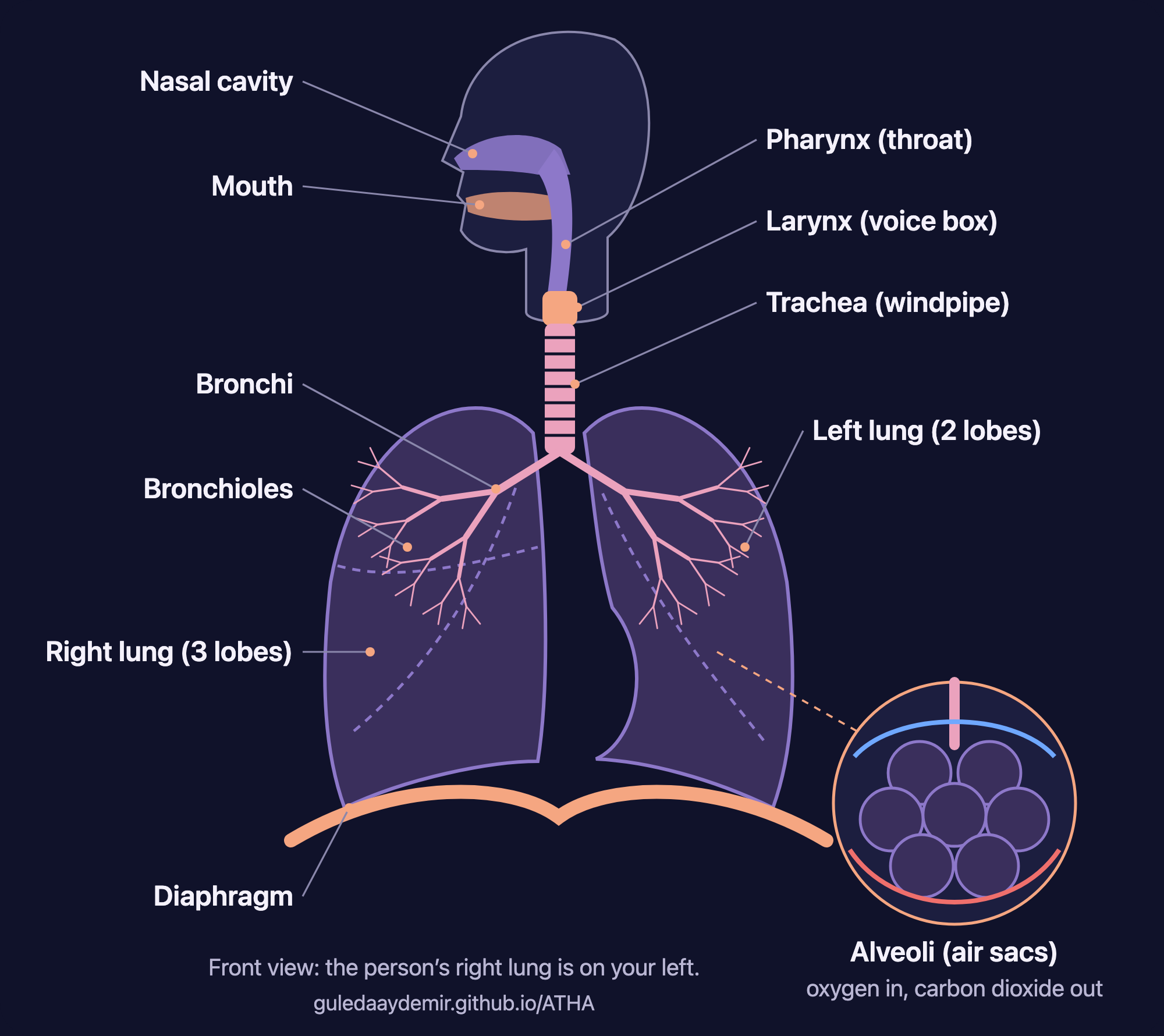
Labelled diagram of the human respiratory system
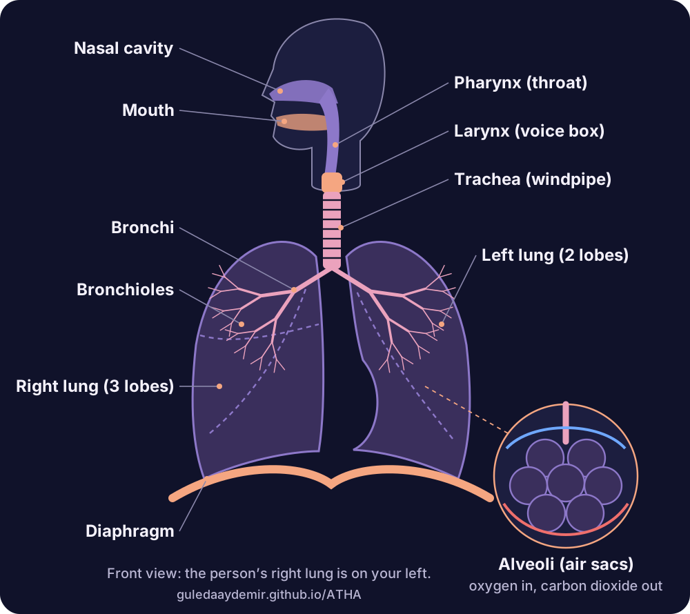
What does each label mean?
- Nasal cavityThe space behind your nose. It warms and moistens incoming air, and tiny hairs called cilia filter out dust.
- MouthThe backup entrance. Air gets in here too, but skips most of the nose’s filtering.
- Pharynx (throat)Shared by your breathing and digestive systems, since it carries both air and food.
- Larynx (voice box)Air passes through here on the way down. On the way out, it’s what makes speech possible.
- Trachea (windpipe)The main tube into the chest, which splits in two at the bottom.
- BronchiThe two big branches, one into each lung.
- BronchiolesThe smaller and smaller branches that fan out through the lung.
- AlveoliTiny air sacs at the very end, wrapped in tiny blood vessels. This is where oxygen goes in and carbon dioxide comes out. You have hundreds of millions of them.
- LungsThe right has three lobes and the left has two.
- DiaphragmThe dome-shaped muscle below the lungs. It moves down as you breathe in and up as you breathe out.
What details do people miss in a respiratory system diagram?
- Left and right are flipped. A diagram is drawn facing you, so the person’s right lung is on your left.
- The lungs don’t match. Three lobes on the right, two on the left, with a dip in the left lung that leaves room for the heart.
- The diaphragm counts. It’s a muscle rather than an airway, but without it, not much air moves.
- The alveoli need a zoom. They’re too small to show at full-body scale, which is why diagrams use a circle close-up.
How do you draw the respiratory system step by step?
- Start with the windpipeA vertical tube down the middle of the page. Add a few rings.
- Split itDraw an upside-down Y at the bottom for the two bronchi.
- Add the lungsTwo tall rounded shapes either side. Leave a dip in the one on your right for the heart.
- Branch the treeKeep splitting the bronchi inside each lung, thinner each time.
- Draw the diaphragmA curved line under both lungs, like a shallow dome.
- Add the head and zoomSketch the nose, mouth and throat at the top, then a circle with a bunch of small sacs for the alveoli.
How can you remember the order?
Say it as a route rather than a list: nose, throat, voice box, windpipe, bronchi, bronchioles, air sacs. Each stop is narrower than the last, and the tree only ever splits. If you lose your place, just ask where the air could go next.
Or do it physically. Take one slow breath, and follow it down in your head as it goes. It’s more memorable than a flashcard, and calmer before a test.
Trace one breath down the diagram
Breathe in for 5 and picture the route. Breathe out for 5 and bring it back.
5 in · 5 out
Follow the orb: in as it grows, out as it shrinks.
Seconds per phase: 5 in · 5 out. Turn on JavaScript to use the timer.
Runs in your browser: nothing is saved or sent. For haptic taps with your eyes closed, or on your wrist, use ATHA on iPhone or Apple Watch.
Common questions
Can I use this respiratory system diagram for my homework or presentation?
Yes, for school work, presentations and non-commercial teaching. Please keep the credit line on the image.
What should a respiratory system diagram label?
At minimum: nasal cavity, mouth, pharynx, larynx, trachea, bronchi, bronchioles, alveoli, lungs and diaphragm.
Why does the right lung appear on the left of the diagram?
Because the diagram shows a person facing you, like a mirror image. Their right side is on your left.
How many lobes do the lungs have?
Five in total: three in the right lung and two in the left.
What is the difference between bronchi and bronchioles?
Bronchi are the larger tubes that branch off the windpipe. Bronchioles are the much smaller branches that carry air on to the alveoli.
Where does ATHA fit in?
I make ATHA, a free breathing app, which is why I care about this drawing more than most people do. Everything in the app is built around that diaphragm at the bottom of the page: slow it down, give it a rhythm, and let your eyes close.

Spot a mistake in the diagram, or teaching with it? I would love to hear about it.
Indie iOS developer, and the maker of ATHA. Say hello on Instagram or at solostaseraofficial@gmail.com.
Sources
Checked on 3 November 2026.
- NHLBI: The respiratory system · Last updated 24 March 2022.
- Cleveland Clinic: Respiratory system · Last updated 9 April 2024.
- KidsHealth (Nemours): Your lungs and respiratory system · Medically reviewed January 2023.
ATHA is a breathing and mindfulness app, not a medical device, and this guide is general information, not medical advice. It doesn’t diagnose or treat any condition. For a symptom that is persistent, severe or worrying, talk to a doctor. In an emergency, call your local emergency number.